Vitamin D: Sources, Forms, Structure, Skin Synthesis & Metabolism
Vitamin D is a fat-soluble secosteroid that plays a crucial role in calcium and phosphate homeostasis, skeletal integrity, and a wide range of extra-skeletal functions. Unlike most vitamins, it can be synthesized endogenously in the skin upon exposure to ultraviolet B (UVB) radiation, making it both a nutrient and a prohormone.
Vitamin D deficiency is a global health concern and is classically associated with rickets in children and osteomalacia in adults. Increasing evidence also links deficiency to osteoporosis, immune dysfunction, cardiovascular disease, and certain cancers.
At the molecular level, vitamin D exerts its effects through the vitamin D receptor (VDR), a nuclear receptor widely distributed across tissues, explaining its diverse physiological roles.
1. Forms of Vitamin D
There are two primary forms relevant to human physiology:
- Vitamin D₂ (Ergocalciferol)
- Derived from plants and fungi
- Produced by UV irradiation of ergosterol
- Present in fortified foods and supplements
- Vitamin D₃ (Cholecalciferol)
- Synthesized in the skin from 7-dehydrocholesterol under UVB radiation
- Also obtained from animal sources (e.g., fatty fish, egg yolk)
- More effective than D₂ in raising serum vitamin D levels
Both forms are biologically inactive and require metabolic activation.
Metabolic Activation and Functional Forms
Vitamin D undergoes two sequential hydroxylation reactions:
- In the liver:
Vitamin D is converted to 25-hydroxyvitamin D [25(OH)D] (calcidiol) - Storage form of vitamin D
- Measured in blood to assess vitamin D status
- Has a relatively long half-life (~2–3 weeks)
- In the kidney:
25(OH)D is further converted to 1,25-dihydroxyvitamin D [1,25(OH)₂D] (calcitriol) - Biologically active form of vitamin D
- Short half-life (~4–6 hours)
- Functions as a hormone regulating calcium and phosphate metabolism
This activation is tightly regulated by parathyroid hormone (PTH), serum calcium and phosphate levels, and fibroblast growth factor-23 (FGF-23).
2. Structure of Vitamin D
Vitamin D is a fat-soluble secosteroid, meaning it is derived from a steroid structure but with a broken B ring (between C9–C10). This structural modification gives the molecule greater flexibility and allows it to function like a hormone by binding to the vitamin D receptor (VDR).
Structurally, vitamin D consists of:
- A modified steroid nucleus (rings A, C, D intact; B ring opened)
- A hydrophobic side chain at carbon 17
- A conjugated triene system (three alternating double bonds), which enables absorption of UVB light and is essential for its synthesis in the skin
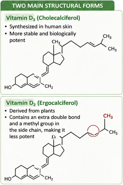
- Vitamin D₃ (cholecalciferol): synthesized in human skin; more stable and biologically potent
- Vitamin D₂ (ergocalciferol): derived from plants; contains an extra double bond and methyl group in the side chain, making it less potent
Vitamin D is highly lipophilic, so it circulates bound to vitamin D-binding protein (DBP) and is stored in adipose tissue.
Biological activity depends on structural modification through hydroxylation:
- 25(OH) Vitamin D (calcidiol): formed in the liver → storage form and the main form measured in blood
- 1,25(OH)₂ Vitamin D (calcitriol): formed in the kidney → active hormonal form
Thus, the structure of vitamin D—particularly its secosteroid nature, side chain, and hydroxylation sites—directly determines its activation, transport, and biological function.
3. Sources of Vitamin D
Vitamin D has a unique feature among vitamins in that it can be synthesized endogenously in the skin. Therefore, its sources are divided into endogenous (primary) and exogenous (dietary) sources.
(a) Endogenous source (Primary source): Sunlight
The most important natural source of vitamin D is cutaneous synthesis following exposure to ultraviolet B (UVB) radiation (290–315 nm).
- In the skin, 7-dehydrocholesterol absorbs UVB radiation
- It is converted to previtamin D₃, which then thermally isomerizes to vitamin D₃ (cholecalciferol)
- This process occurs mainly in the epidermis (stratum basale and stratum spinosum)
For most individuals, sunlight is the dominant source of vitamin D, and dietary intake alone is often insufficient.
(b) Animal sources (Vitamin D₃ / cholecalciferol)
Vitamin D₃ is the primary form found in animal-derived foods:
- Fatty fish (e.g., salmon, mackerel, sardines, tuna) – among the richest natural sources
- Cod liver oil – historically one of the most concentrated sources
- Egg yolk – variable content depending on hen diet and sun exposure
- Liver and other organ meats – minor amounts
- Fortified dairy products (in many countries) – significant dietary contributor where fortification is practiced
(c) Plant and fungal sources (Vitamin D₂ / ergocalciferol)
Vitamin D₂ is primarily obtained from UV-exposed plant and fungal sources:
- UV-irradiated mushrooms (contain ergosterol → ergocalciferol)
- Yeasts and fungi after ultraviolet exposure
- Naturally occurring plant foods contain very little to no vitamin D unless UV-treated
(d) Human milk and other considerations
- Human breast milk contains low levels of vitamin D, even in vitamin D–sufficient mothers
- Therefore, infant supplementation is often required, especially in exclusively breastfed infants
- Vitamin D content in milk and eggs varies significantly, depending on:
- Animal diet
- Sunlight exposure
- Feed fortification practices
4. Photochemical Regulation and Photoproducts
The formation of vitamin D from ergosterol and 7-dehydrocholesterol is not fully efficient. Several photoproducts are formed simultaneously, including:
- Lumisterol
- Tachysterol
These compounds are biologically inactive and form part of a natural regulatory mechanism called the photostationary system, which prevents excessive vitamin D production during prolonged sunlight exposure by shifting photochemical equilibrium.
Tachysterol can be hydrogenated to form dihydrotachysterol (AT-10 D₃), a synthetic analog with vitamin D–like activity, particularly in calcium metabolism.
5. Transport and Activation
Vitamin D is a lipid-soluble molecule and does not circulate freely in plasma. After synthesis in skin or absorption from diet, it binds to vitamin D–binding protein (DBP) for transport to the liver.
Vitamin D exists in biologically inactive forms and requires two hydroxylation steps for activation:
- Liver: Conversion to 25-hydroxyvitamin D (calcidiol)
- Kidney (proximal tubules): Conversion to 1,25-dihydroxyvitamin D (calcitriol)
Calcitriol is the biologically active form responsible for most physiological actions.
6. Endocrine Regulation of Vitamin D Metabolism
Vitamin D activation is tightly regulated by endocrine signals:
- Parathyroid hormone (PTH): stimulates renal 1α-hydroxylase → increases calcitriol
- Low serum calcium: indirectly increases calcitriol via PTH
- High phosphate levels: stimulate FGF-23
- Fibroblast growth factor-23 (FGF-23): inhibits calcitriol synthesis and promotes phosphate excretion
This integrated axis maintains calcium and phosphate balance within narrow physiological limits.
Calcitriol also participates in negative feedback inhibition of PTH, forming a self-regulating endocrine loop.
7. Vitamin D Receptor (VDR) and Genomic Action
The biological actions of vitamin D are mediated through the vitamin D receptor (VDR), a nuclear receptor present in target tissues such as intestine, bone, kidney, and immune cells.
Calcitriol binds to VDR and regulates gene transcription, leading to synthesis of proteins involved in calcium transport, bone remodeling, and immune regulation. This explains why vitamin D is now considered a hormone rather than a simple vitamin.
8. Storage, Distribution, and Half-life
Vitamin D is stored primarily in:
- Adipose tissue
- Liver (as 25-hydroxyvitamin D)
Because of its fat solubility, it has a long biological half-life, allowing maintenance of plasma levels over extended periods even with intermittent intake or sunlight exposure.
9. Regulation of Cutaneous Synthesis
Cutaneous synthesis of vitamin D is influenced by several physiological and environmental factors.
- Melanin acts as a natural sunscreen by absorbing UVB radiation and is the most important biological inhibitor of vitamin D synthesis.
- Keratin provides a minor physical barrier.
- Therefore, individuals with darker skin pigmentation require longer sunlight exposure to produce equivalent vitamin D levels.
- Aging reduces 7-dehydrocholesterol content in skin, decreasing synthesis.
- Obesity reduces bioavailability due to sequestration in adipose tissue.
- Environmental factors such as latitude, season, time of day, clothing, sunscreen use, and air pollution significantly influence production.
Importantly, UVB radiation is completely blocked by ordinary window glass and significantly reduced by atmospheric pollution, smoke, and dust.
10. Food Fortification and Modern Sources
Modern nutrition utilizes UV irradiation and fortification techniques to increase vitamin D content in foods, especially:
- Milk products
- Mushrooms
- Cereals and fortified foods
This compensates for limited natural dietary sources.
11. Vitamin D as a Hormone Precursor
It is now well established that vitamin D is not merely a dietary vitamin but a hormone precursor synthesized in the skin under environmental regulation. Its activation is tightly controlled by endocrine systems involving the kidney, parathyroid glands, and liver.
12. Clinical Correlation
Defects in vitamin D synthesis or action lead to:
- Rickets (children): defective mineralization of growing bone
- Osteomalacia (adults): defective mineralization of mature bone
These conditions reflect failure of calcium–phosphate homeostasis due to inadequate calcitriol activity.