Effects of Deficiency of Vitamin C
1. Functions of Vitamin C
Vitamin C is concerned fundamentally with the formation of intercellular substances, including collagen of fibrous tissue structures, the matrices of bone, cartilage and dentine, and all non-epithelial intercellular cement substances, including that of vascular endothelium. It acts as an essential cofactor for the hydroxylation of proline and lysine residues in collagen synthesis, which is necessary for stable triple-helix formation of collagen. The connective tissue has the highest concentration of vitamin C and after wounds it accumulates quickly in the scar tissue. An excessive intake of vitamin C is claimed to decrease the incidence of atherosclerosis and the common cold, although current evidence for these effects remains limited and not conclusively established.
Vitamin C (ascorbic acid) plays a central biochemical role in maintaining the integrity of connective tissue. Its most critical function is enabling the post-translational modification of collagen, where it ensures proper hydroxylation of amino acids. Without this step, collagen fibers become unstable and weak, leading to fragile tissues throughout the body. Because collagen is the structural framework of skin, blood vessels, cartilage, and bone, vitamin C indirectly influences wound healing, vascular strength, and skeletal development. Its accumulation in healing tissue reflects its importance in tissue repair processes.
2. Disease Due to Deficiency
In the deficiency of vitamin C, the disease called scurvy (scorbutus) results. Scurvy is liable to occur in artificially-fed (formula-fed) infants. Breast-fed infants rarely suffer from scurvy. It should be noted that subclinical scurvy occurs much more frequently than frank scurvy. Scurvy has the following clinical picture:
Scurvy represents a systemic breakdown of connective tissue integrity due to failure of collagen synthesis. It develops gradually, often beginning with subtle biochemical deficiency before clinical signs appear. Infants dependent on formula lacking adequate vitamin C are particularly vulnerable, while breast milk generally provides sufficient levels. Subclinical deficiency is common and may go unnoticed until stress or illness reveals overt symptoms.
3. Clinical Features of Scurvy
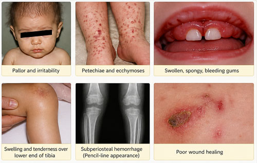
3.1 Hemorrhagic manifestations
- Hemorrhages from the mucous membranes of the nose, mouth and gastrointestinal tract, skin, muscles, and in the subperiosteal tissues.
- The most frequent site of lesions is the gums, which show swelling, redness, ulceration, etc., which may lead to gangrene and loss of teeth.
- Capillaries show increased fragility.
- Bleeding may also present as petechiae, purpura, ecchymoses, and generalized capillary oozing.
The hallmark of scurvy is bleeding due to weakened capillary walls. Since collagen is essential for vascular support, deficiency leads to fragile blood vessels that rupture easily under minor stress. Gingival tissues are especially affected because of their high vascularity and constant mechanical irritation.
3.2 Systemic Symptoms
- Anemia, weakness and emaciation; there is a tendency to remain motionless.
- Fatigue, irritability, and general malaise are also common.
- Vitamin C deficiency may also worsen iron-deficiency anemia due to impaired iron absorption.
Systemic symptoms reflect both nutritional impairment and chronic micro-hemorrhages. Vitamin C enhances iron absorption in the intestine; thus, its deficiency compounds anemia. Patients often appear lethargic due to reduced oxygen delivery and metabolic inefficiency.
3.3 Skin Manifestations
- Skin shows discoloration of perifollicular areas, especially over the back and thighs.
- “Corkscrew hairs” due to follicular hyperkeratosis are often seen.
Skin changes result from defective connective tissue around hair follicles. The characteristic corkscrew hairs occur because hair shafts grow without proper structural support in the dermis, leading to coiling and distortion.
3.4 Healing Defects
Since vitamin C is necessary for wound healing, its deficiency results in delayed healing of wounds and fractures.
Wound healing depends heavily on collagen deposition. Without vitamin C, fibroblasts cannot form stable connective tissue, resulting in prolonged recovery times and weak scar formation.
3.5 Skeletal and dental changes
At the growing ends of long bones, osteoblastic function is arrested, but there is continued calcification of cartilage which leads to a piling up of calcified matrix. This is brittle and commonly shows fragmentation, while fractures may take place through the rarefied metaphysis immediately adjacent. In addition, a fibrous union is formed between the diaphysis and the epiphysis, which may be seen as an enlargement of the costochondral junctions. Characteristic hemorrhages occur beneath the periosteum of long bones; the hematomas may become calcified. There is pain in joints. Acute tenderness of the limbs causes the infant to cry with pain when limbs are touched. The legs are held in “the frog position” in which the thighs are abducted and semi-flexed. X-rays show the heavy “white line” of calcified matrix at the ends of long bones, the rarefied zone adjacent to it and, in severe cases, a ground-glass appearance of the long bones and epiphyses due to generalized rarefaction. Tooth development is impaired in the very young; this is due to non-formation of dentine by odontoblasts.
Skeletal manifestations are especially severe in growing children because bone formation is highly active. The imbalance between cartilage formation and defective bone matrix leads to structural weakness, fractures, and deformities. Subperiosteal hemorrhages contribute significantly to pain and tenderness, while radiological findings reflect disrupted bone growth and mineralization. Dental defects arise from failure of odontoblasts to produce dentine, resulting in weak and poorly formed teeth.
4. Immune System Effects
There may also be impaired immunity with increased susceptibility to infections due to defective leukocyte function.
Vitamin C supports leukocyte activity, including chemotaxis and phagocytosis. Deficiency weakens immune defenses, making patients more prone to bacterial and viral infections and prolonging recovery from illness.
5. Diagnosis of scurvy
The diagnosis of scurvy is made from the patient’s history, physical findings and the following laboratory investigations:
Diagnosis is based on a combination of clinical suspicion and biochemical confirmation. Dietary history is particularly important, especially in infants or malnourished individuals.
5.1 Capillary fragility test (tourniquet test)
Capillary fragility test or tourniquet test: A blood pressure cuff is maintained in place while it is inflated to a pressure between systolic and diastolic pressure for 5 to 15 minutes and the number of petechial hemorrhages in a 2.5 cm diameter circle on the upper forearm is recorded. A positive test (petechiae more than 10) is evidence of a platelet or vascular defect. This test is however not specific for vitamin C deficiency.
This test assesses capillary integrity. However, because many disorders can increase bleeding tendency, it is supportive rather than definitive for scurvy.
5.2 Plasma ascorbic acid
Determination of plasma ascorbic acid: Normal plasma ascorbic acid level is 0.7 to 1.2 mg%; a level below 0.4 mg% indicates severe deficiency.
Plasma vitamin C measurement provides a direct biochemical estimate of deficiency, though it may fluctuate with recent dietary intake.
5.3 Buffy coat vitamin C level
The ascorbic acid level in the white cell–platelet (buffy coat) layer is more significant. A level below 4 mg per 100 mL is closely correlated with scurvy (normal = 25 to 30 mg per 100 mL).
White blood cells store vitamin C more reliably than plasma, making this measurement a more accurate indicator of tissue stores.
5.4 Saturation (load) test
Saturation or load test: on giving a standard dose of ascorbic acid to the patient, very little of it appears in the urine. This is because the tissues, being deficient in ascorbic acid, retain an excess of it. It is only when tissues become saturated with ascorbic acid that it starts appearing in urine in appreciable amounts.
This test evaluates tissue depletion. In deficiency states, administered vitamin C is rapidly absorbed and retained until body stores are replenished.
6. Time course of deficiency
The half-life of ascorbic acid is about 16 days and it takes 3 to 4 months for scurvy to develop if a person is put on a diet lacking vitamin C.
Because vitamin C is water-soluble and not extensively stored, deficiency develops relatively quickly compared to fat-soluble vitamins. Clinical scurvy typically appears after several months of inadequate intake, once tissue reserves are exhausted.